

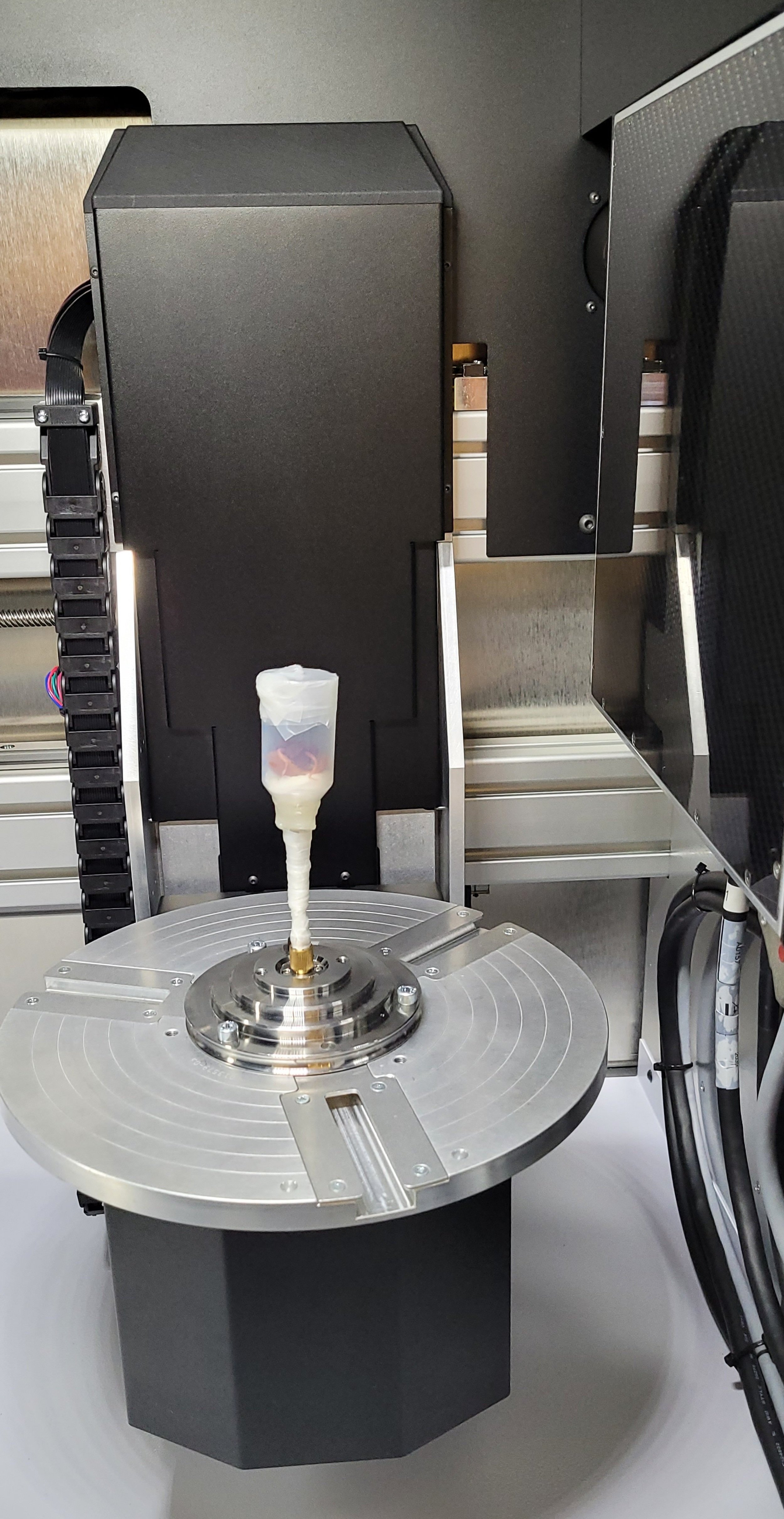
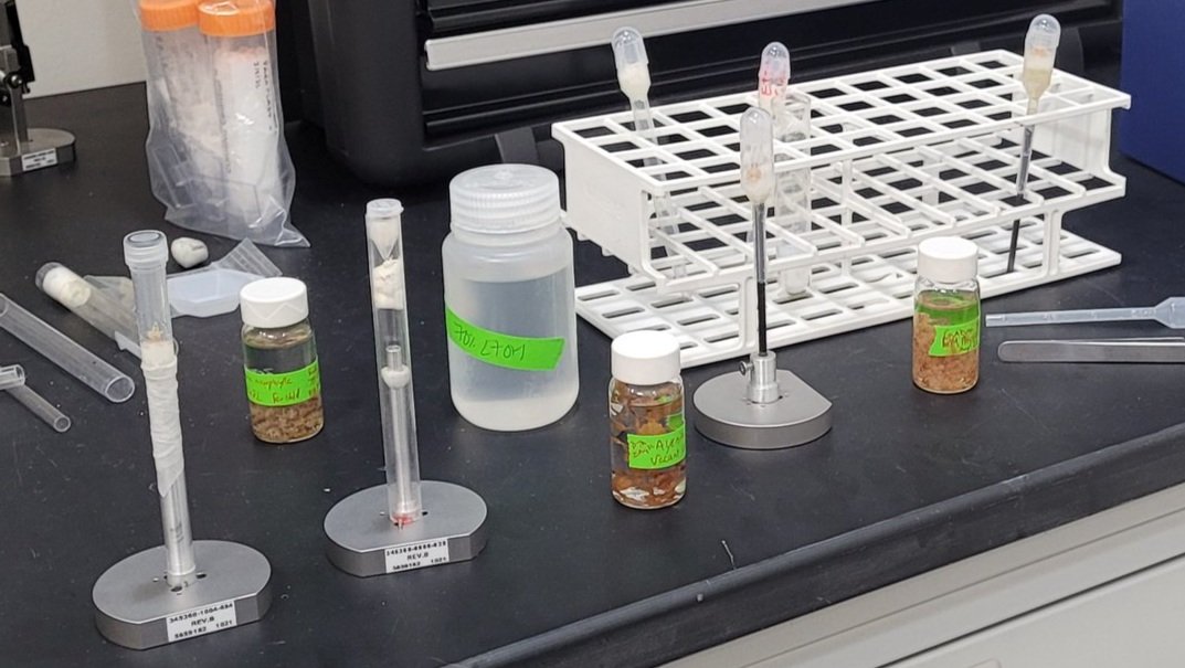
Most people don’t realize how much crafting goes into CT scanning. A lot of the times, I am scrambling for any appropriately sized vessels around lab. Then I cut them up and hot glue them together.
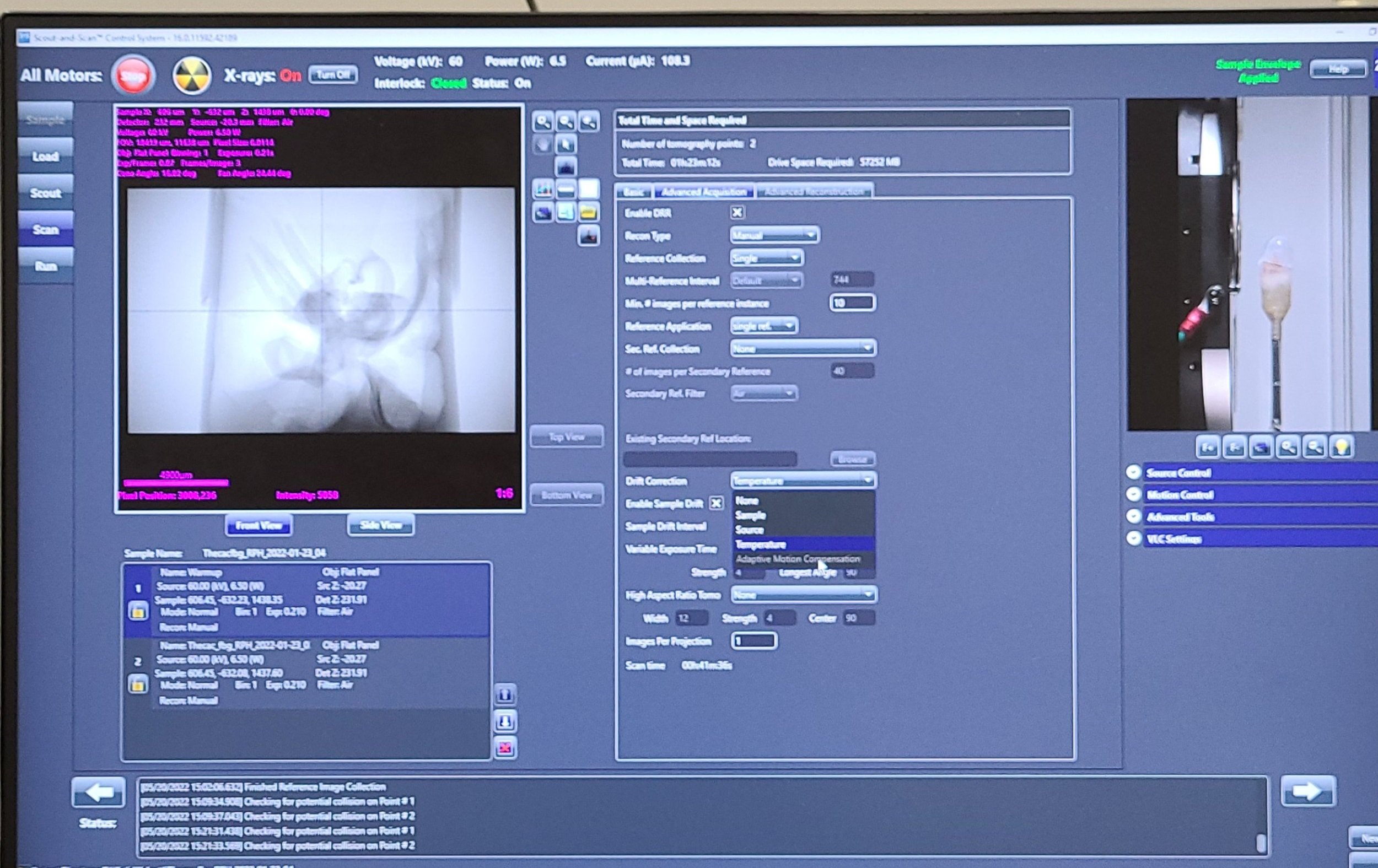
There’s an X-ray preview on the left and a camera view of the specimen in the scanner on the right.

Using a DSLR Camera and a rotating stage to take images of a flower in 360 degrees from multiple angles. Images will be stitched together as an accurate 3D surface model.
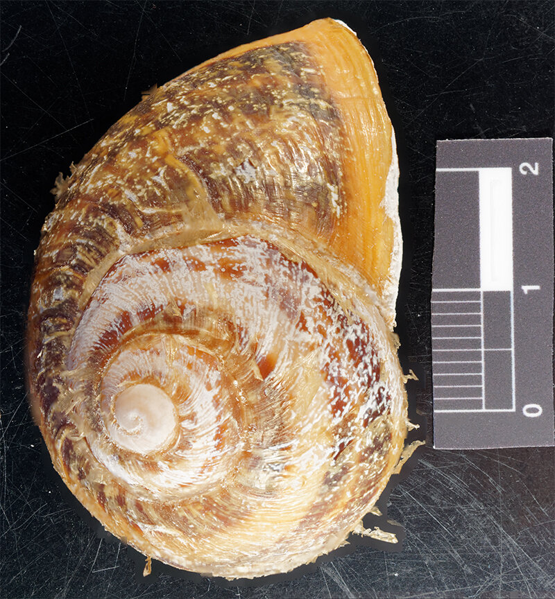
To determine which species of snail we were working with to develop HAPI Helix, we used a combination of DNA barcoding and shell morphology.

So organs could be seen in their natural positions, we used micro-CT scans from whole snails to make HAPI Helix digital 3D model. In order to increase absorption of the iodine-based staining solution, I used a Dremel to drill slits into the snail shell.

This sea orange (Tethya aurantium) is actually a marine sponge, and it was found while I was on a marine biology excursion on the Slovenian coast. We did a dredge off of a 10-person motorboat to collect sponges, tunicates, hermit crabs, and other benthic marine life. There are also sea lemons (Tethya citrina), and they are easy to tell apart from sea oranges. They are usually a little bit smaller, more yellow, and they don't have the radially-oriented long, spikey things (spicules) that you can see here. Take a look at the next photo to see the other kind of spicules that sea oranges have!

Spicules are used as a sort of skeleton to provide support to sponges. Their shape varies a lot within sponges, they can be identified based on whether they are made out of calcium carbonate (like chalk), silicon dioxide (like glass), or if there are no spicules and a "spongey" web (spongin protein) is present instead. After determining this main difference, you can look closer to the shapes of the spicules for help with identification to family, genus, or species. While the Sea Orange also has long, slender spicules that are visible with the naked eye in the previous photo, they also have these small star-shaped spicules (shown at 40x magnification).

For a short research project, I am studying the foot morphology of Sea Spiders (Class: Pycnogonida) and relating the differences in the number/size/location of spikes to differences observed in DNA barcoding data. Not fully true to their name, they belong to the same taxa, Chelicerata, which is nestled within Arthropoda. Chelicerata also contains spiders and horseshoe crabs. As they are mostly benthic (live at the bottom of the sea, as opposed to free-swimming), their feet are very important for mobility, feeding, mating, and even for glueing their offspring to clams and polychaete worms.
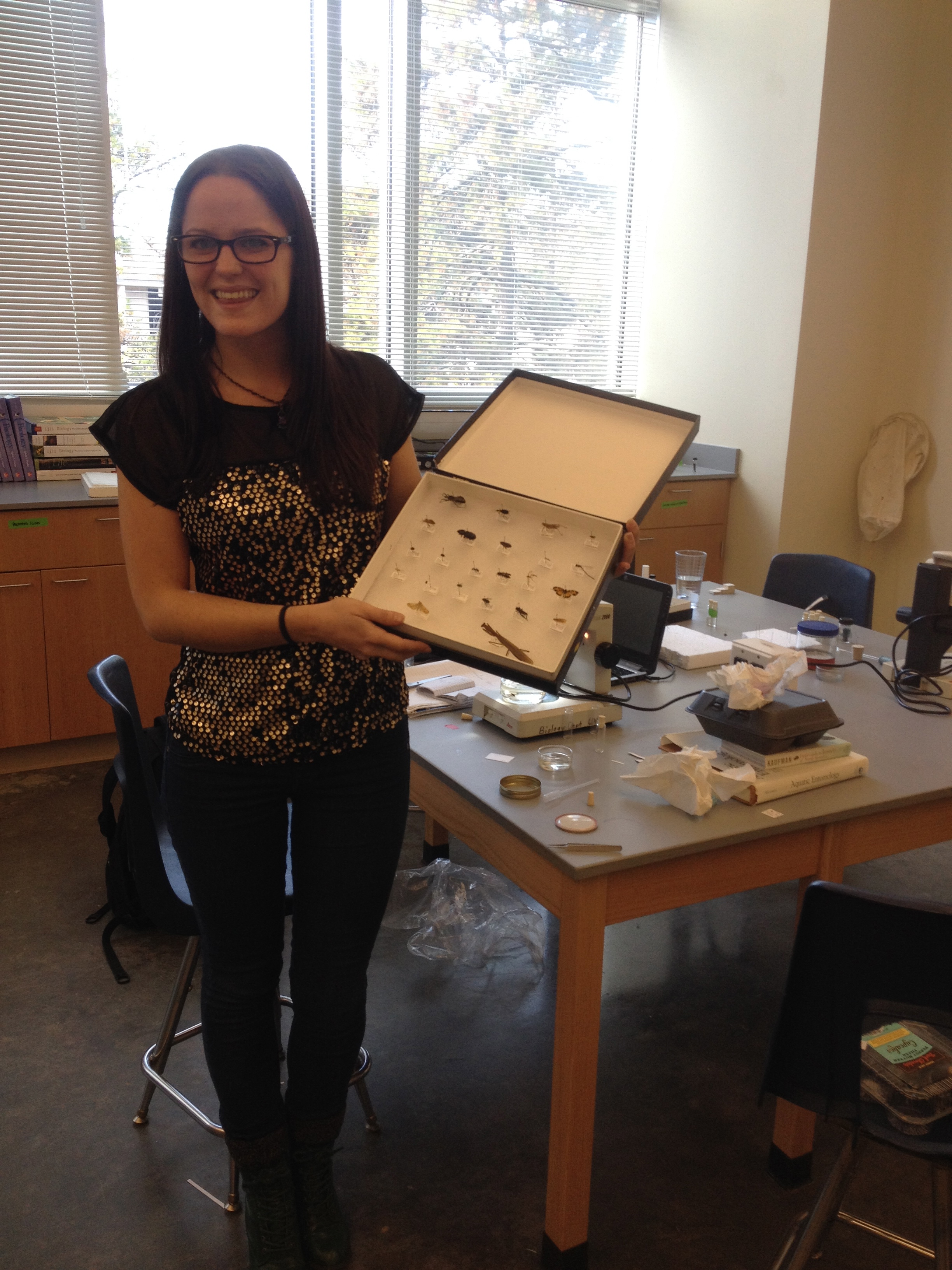
During my last few weeks of insanity before finishing my Bachelor's degree. So happy to have finished my last biological collection as an undergrad, but looking forward to more collecting and identifying in the future.
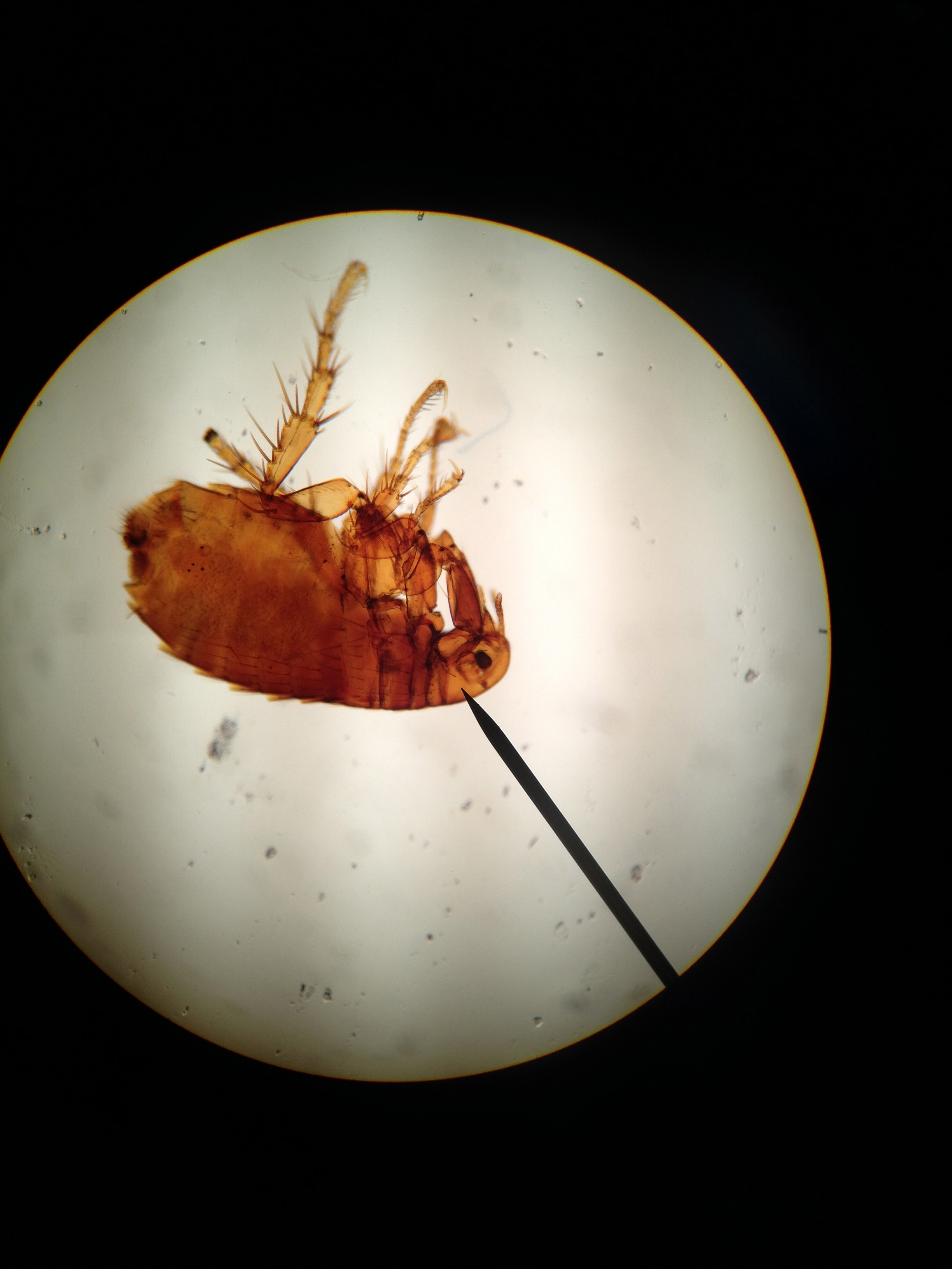
Fleas are ectoparasites of mammals, and their most posterior set of legs contain pads of the most efficient elastic protein known: resilin. This protein is responsible for the fast wing speeds of flies, and the ability of fleas to jump so high. Fleas also carry black plague, which is caused by the bacteria Yersinia pestis. The hypothesized coevolution of this bacteria from water-dispersal to flea-dispersal is a pretty cool story I researched a bit last fall. I will write a blog post about it soon.
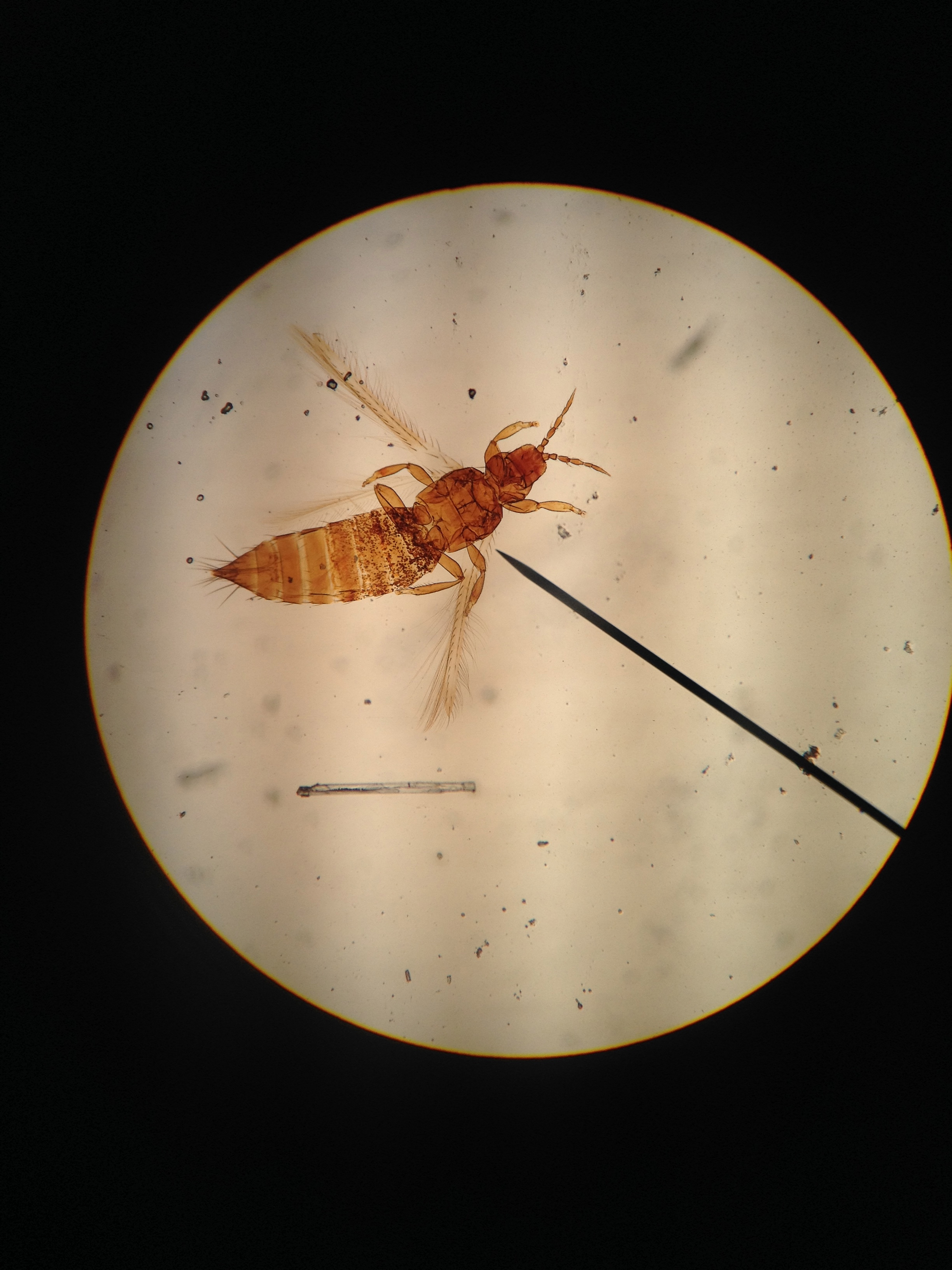
Thysanopterans are a group of hemimetabolous insects characterized by asymmetrical mouthparts. One mandible is vestigial, while the other is enlarged and modified for piercing into tissue for consumption. Hemimetabolous insects are characterized by incomplete metamorphosis though life stages egg, larva, and adult. In addition to these three stages, holometabolous insects undergo a quiescent pupa stage before reaching adulthood.
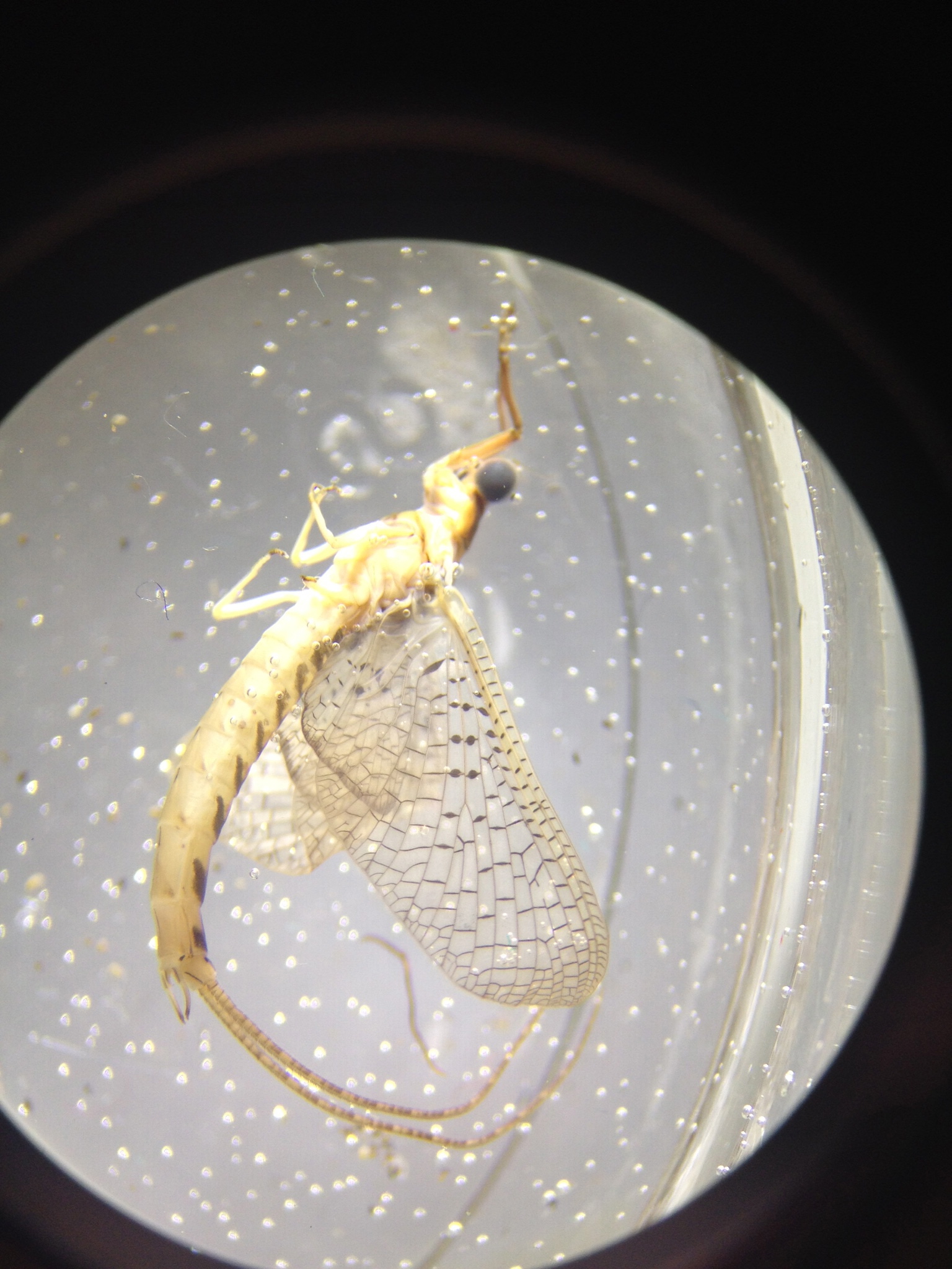
This is an adult mayfly in the family Ephemeridae. Typically, insect families end in -idae, while plant families end in -aceae. The name "Ephemeroptera" is derived from the Greek "ephemera" relating to the short lifespan of the adult mayfly. Adults have either vestigial or no mouthparts, since they do not need to feed after their larval stage.
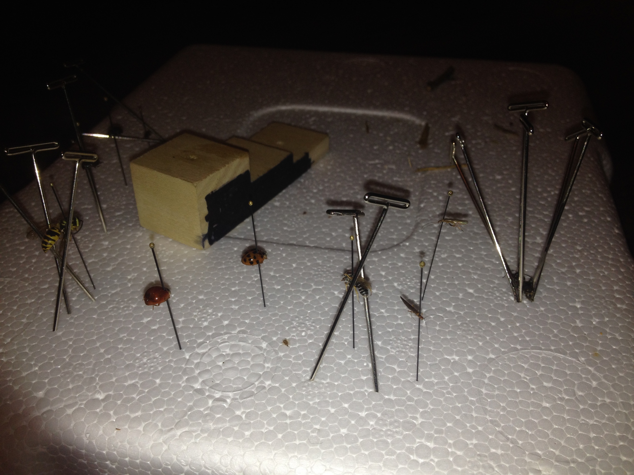
When pinning insects for collection, you select one pin of appropriate thickness and usually place it slightly to the right of their body's midline. Once the body is perpendicular with the pin, you place the insect on the positioning block so all specimens are at the same height. Pinned specimens are stabilized in styrofoam, and thicker positioning pins are used to stabilize various body parts for a few days before being placed in a more permanent place for storage and identification.
Before germination of Acacia saligna, seeds were scarified and the elaiosome was removed. An elaiosome is hypothesized to be a nutritive reward associated with ant dispersal, and its presence or absence does not have a known impact on plant fitness.
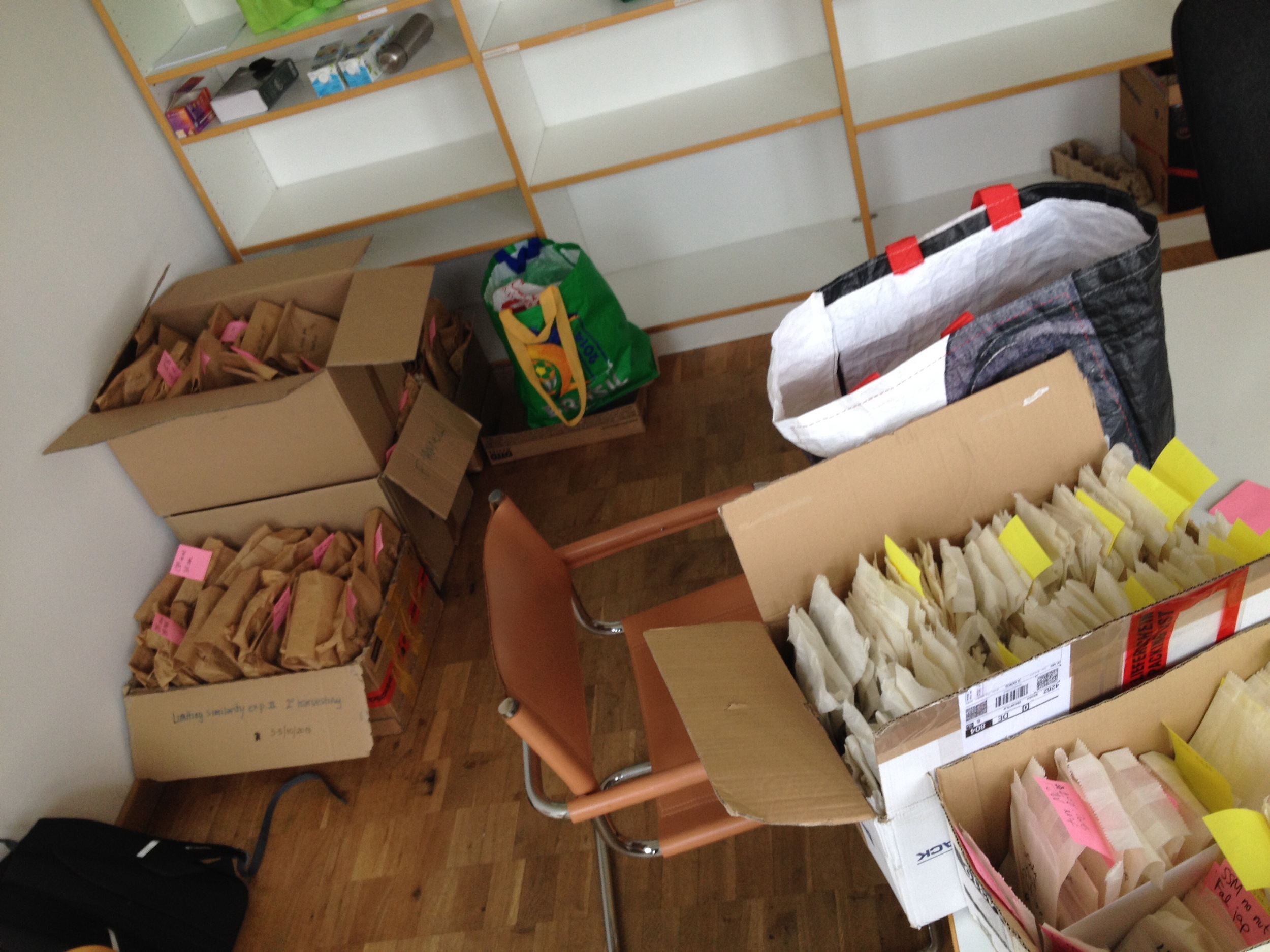
After harvesting the native community experiment, labeled biomass samples were oven-dried over the weekend and sorted into boxes. Before massing samples, they were stored in the drying oven again to eliminate any extra water weight that may have accumulated from humidity in the air.

Before starting an experiment using live plants, germination trials are performed to know how successful seeds are germinating, and to determine how many seeds will be needed for each trial.
Seeds are put into chambers at designated light and temperature intervals (ie 12 hours light/dark at 20/10°C). Currently, we're looking at Festuca rubra (a grass), Daucus carota (also known as carrots!), and Olea europaea (olives).

Counting the number of seeds germinated per petri dish. We will do this a few times over the course of several days to get an average and representative germination rate for the species studied.

Scarifying some Cytisus scoparius (L.) Link seeds from Norway using a scalpel and forceps.
Some species require some sort of stimulus to break dormancy. Usually, this is either scarification (mechanical or chemical) or stratification (enduring periods of cold temperatures). These stimuli mimic natural triggers to seed germination: passing through the digestive tract of a dispersal agent (mammal, bird, etc) or enduring winter before the spring growing season begins.

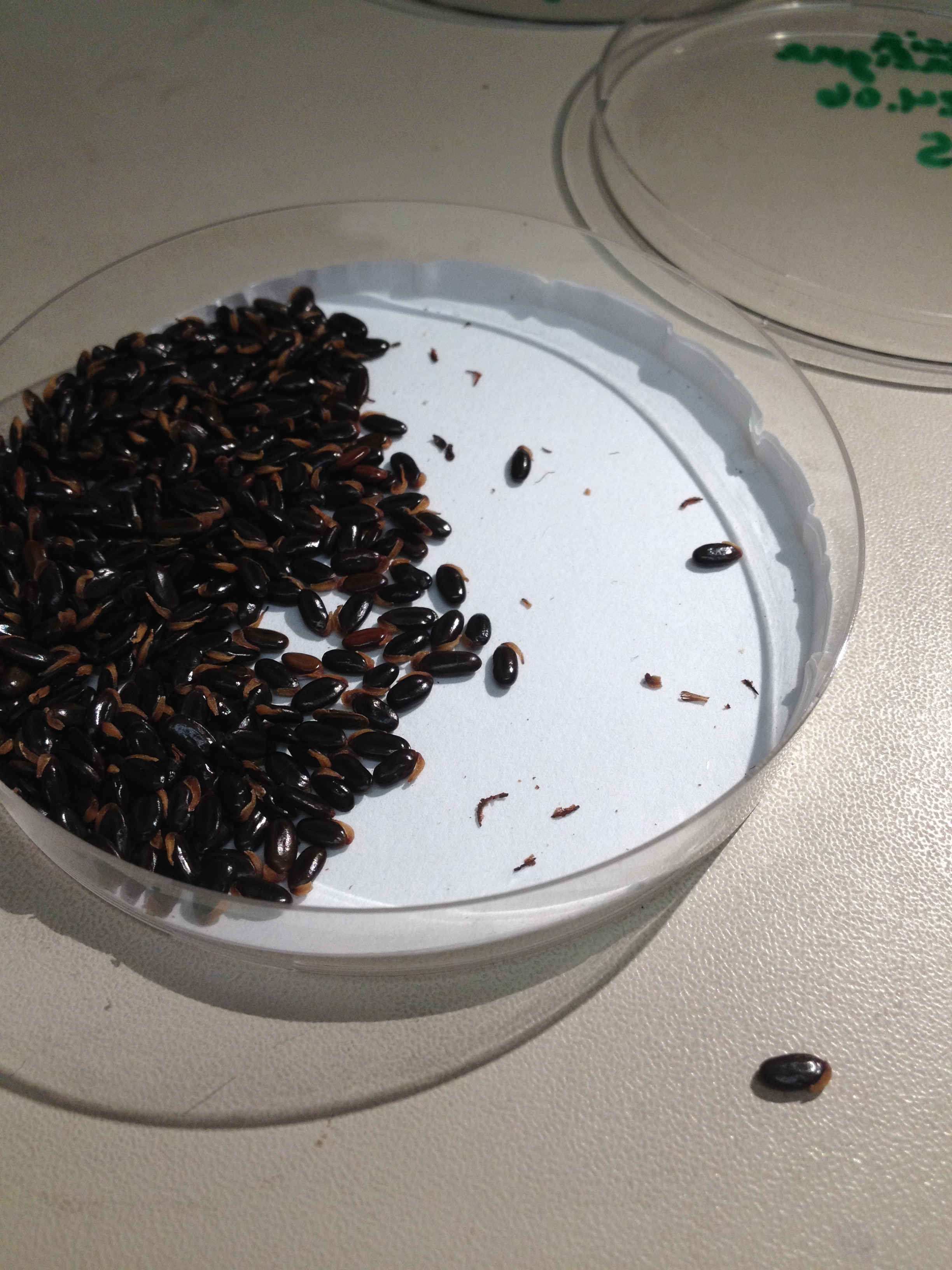
Massing out some seeds for a community ecology experiment in the greenhouse.

A closeup of the excitement that is EXPERIMENT PREP

Although it looks like Marchantia, I am not certain to what genus this little liverwort belongs. I do know that it is currently undergoing asexual reproduction, because of the presence of gemmae cups on its "leaves." Since liverworts do not have true vasculature, and the leaflike structures are analagous, not homologous, to true leaves.

The soil gym, as I more affectionately call it. To process frozen soil samples collected around Bavaria, we must break up samples using any variety of graceful techniques. I like to pretend that I'm working with pizza dough, as I throw the bag of frozen soil onto the counter, rotate, and repeat.

When I need to brush up on my Asteraceae morphology, I use this study guide I created of Rudbeckia laciniata L.
1) Disc flower
2) Receptacle
3) Phyllary (involucral bract)
4) Synantherous stamens (connate at anthers)
5) Ray flower
6) Chaff
Note that there is no pappus

40 of my best specimens collected Fall 2013, preserved in vials filled with alcohol

The larval stage of Utterbackia imbecillis (Say, 1829), a bivalve native to western North Carolina in the family Unionidae. It is parasitic on fish gills for transportation and gas exchange.

The lophophore (U-shaped feeding structure) of Pectinatella magnifica (Leidy, 1851), a freshwater Bryozoan.

The home of my plant collections from Spring, Summer, and Fall 2013. I am not sure how many hours I spent referencing specimens in this room.