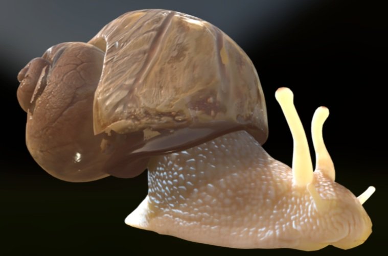
HAPI Helix Educational 3D Model

Photo taken by Bernhard Ruthensteiner, 7 March 2018 at Klenze Gymnasium Munich.
For my Master's thesis project, I created a digital 3D model of a Roman snail (Helix pomatia, also known as an edible or Burgundy snail). The model was called HAPI Helix - Harm free anatomy and physiology instruction for Helix pomatia. After HAPI Helix was completed, I used it to teach students alongside traditional dissection and then measured learning of 8th grade biology students who used dissection only, HAPI Helix only, and HAPI Helix + dissection.
To create HAPI Helix, we used micro-CT scans to generate the model in Amira software. You can see the basic workflow below.
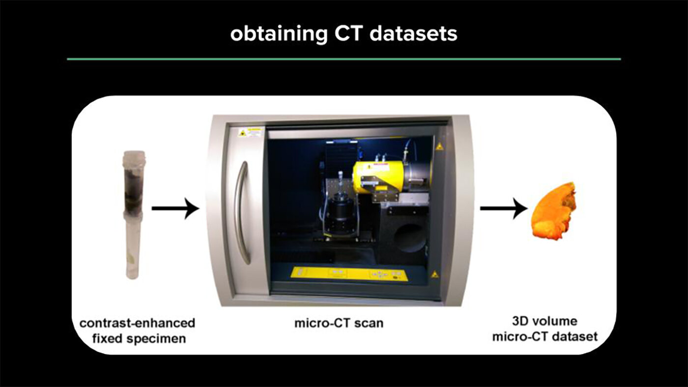
Slide 1: To obtain CT datasets from a specimen (a snail’s heart and kidney are shown), the specimen’s position was fixed in a closed contained to ensure it doesn’t move or dry out during the scan. Then the specimen is scanned, which results in a CT dataset consisting of X-ray image series that can be viewed in 3-dimensions as virtual cross sections.
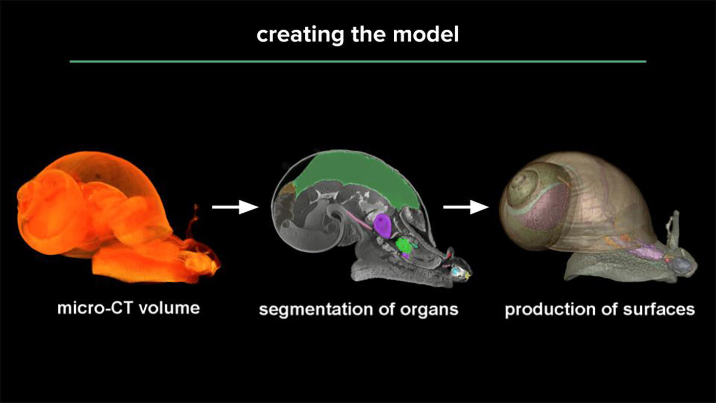
Slide 2: To create a digital 3D model using CT scans, these virtual cross sections are viewed in Amira Software, and then trace the outlines of different organs to create an anatomically-realistic digital 3D model.
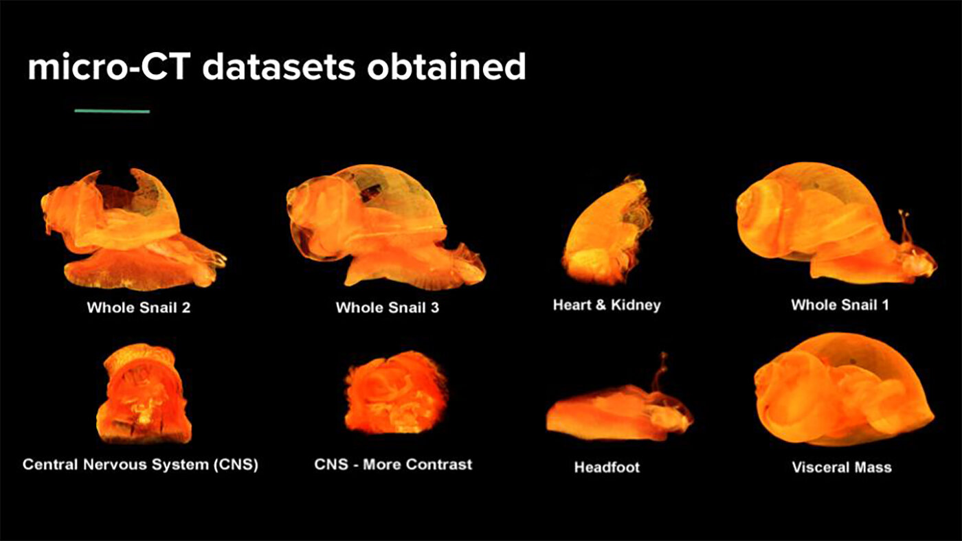
Slide 3: Sometimes multiple scans of body regions are needed to get better resolution and make a more detailed model. All CT datasets obtained to create HAPI Helix are shown here.
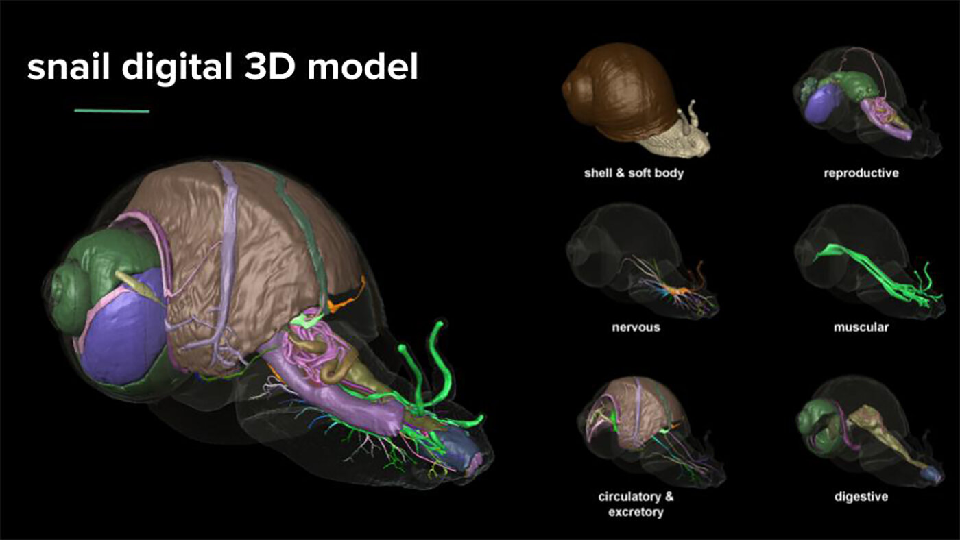
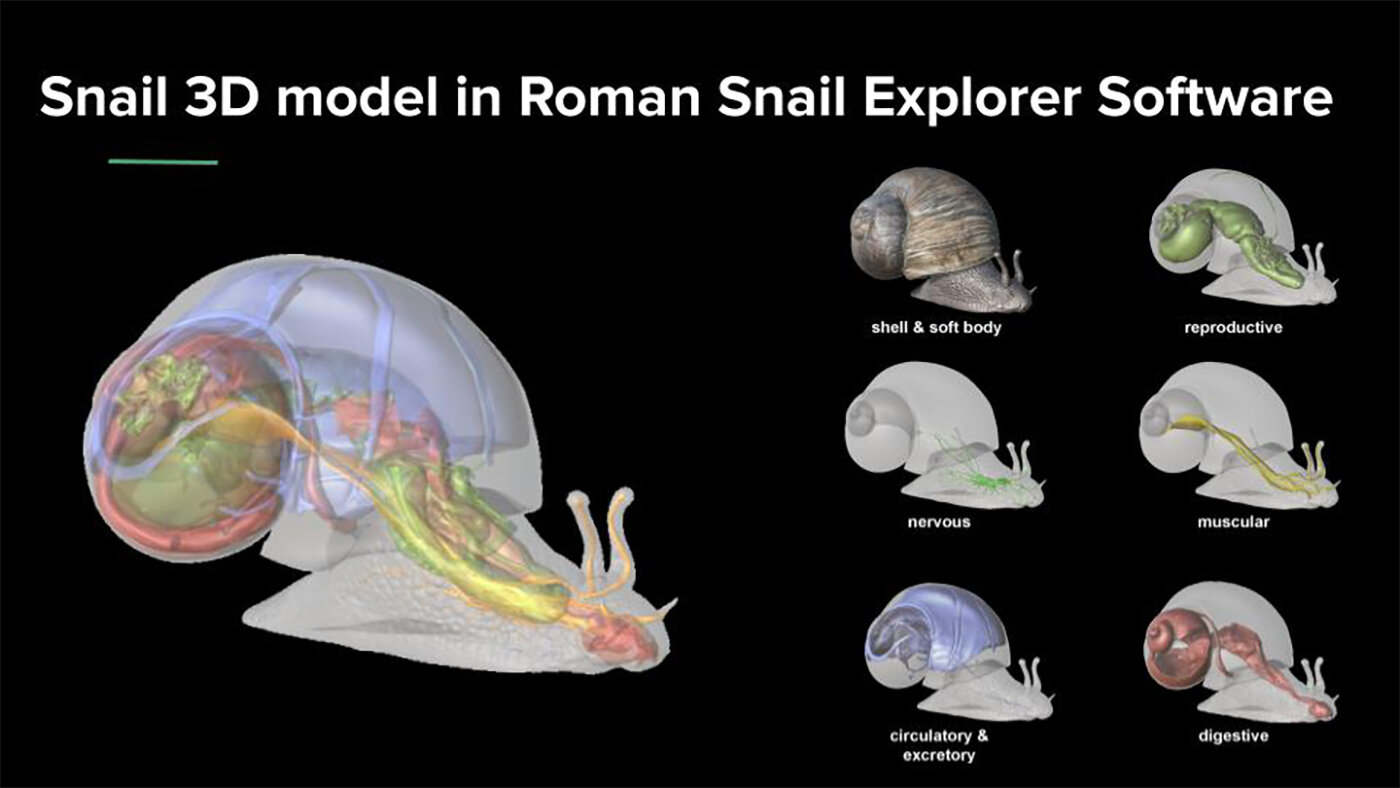
Slide 4: Final 3D surface models of all body systems of Helix pomatia.
After creating HAPI Helix, we began a partnership with a Biological 3D Imaging Company,
Effigos, AG (Leipzig), to create Roman Snail Explorer 3D Educational Software.
Slide 1: Models for all body systems were simplified and embedded in Roman Snail Explorer Software.
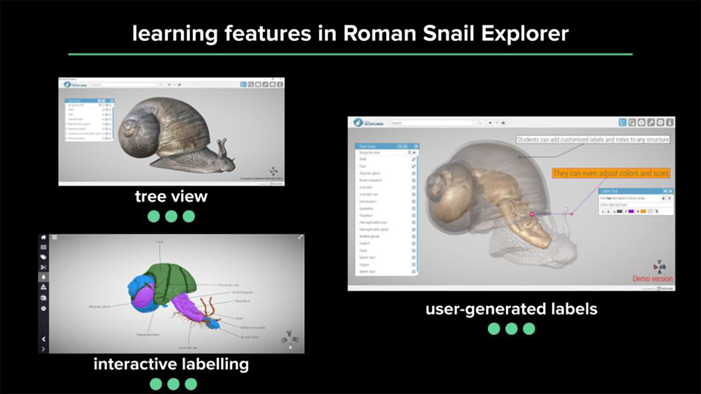
Slide 2: The software is available for desktop, tablets and web browsers and includes learning features like a tree view of anatomical structures that can be toggled on/semi-transparent/off, interactive labeling that changes position with model rotation, and the option to your own labels to refresh your memory during lesson review.
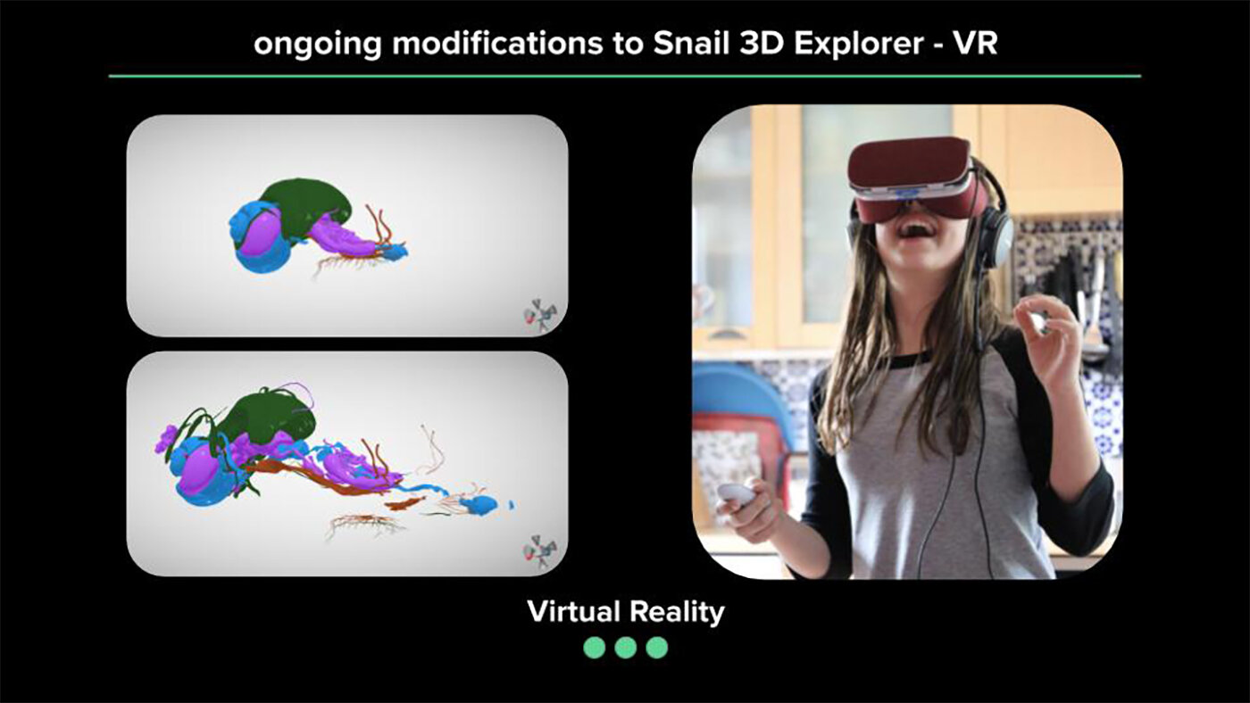
Slide 3: Snail Explorer Software is also available in Virtual Reality for Samsung Oculus. The first time I used it, I pulled apart body systems in 3D space and was thrilled to be surrounded by floating, spinning snail organs. Photo on right: "Virtual joy" by Jonathan Tallon is licensed under CC BY-NC-SA 2.0
Once Roman Snail Explorer Software was developed,
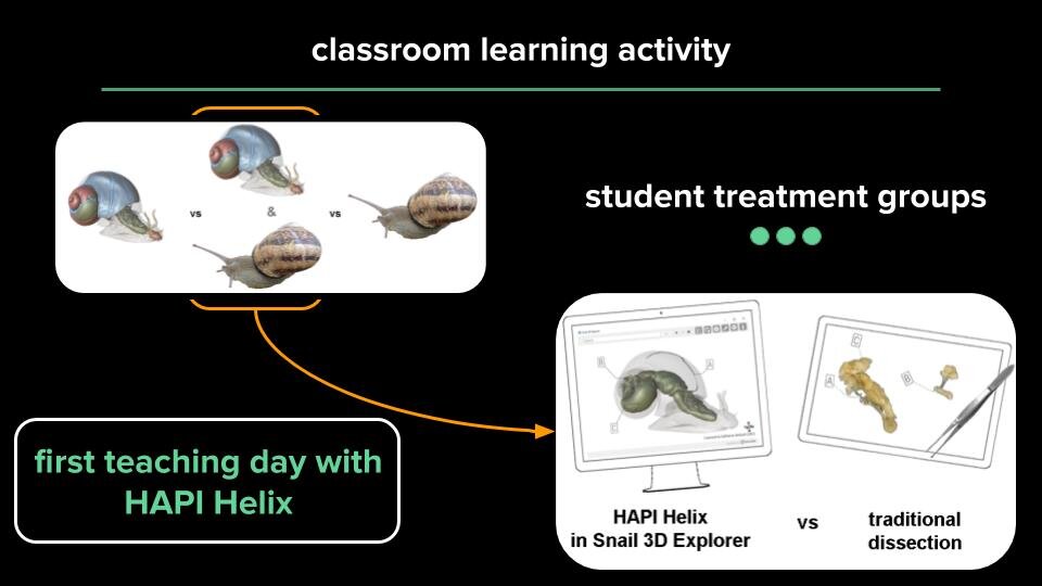
we brought it into an 8th grade biology classroom to measure student learning
using HAPI Helix + traditional dissection.
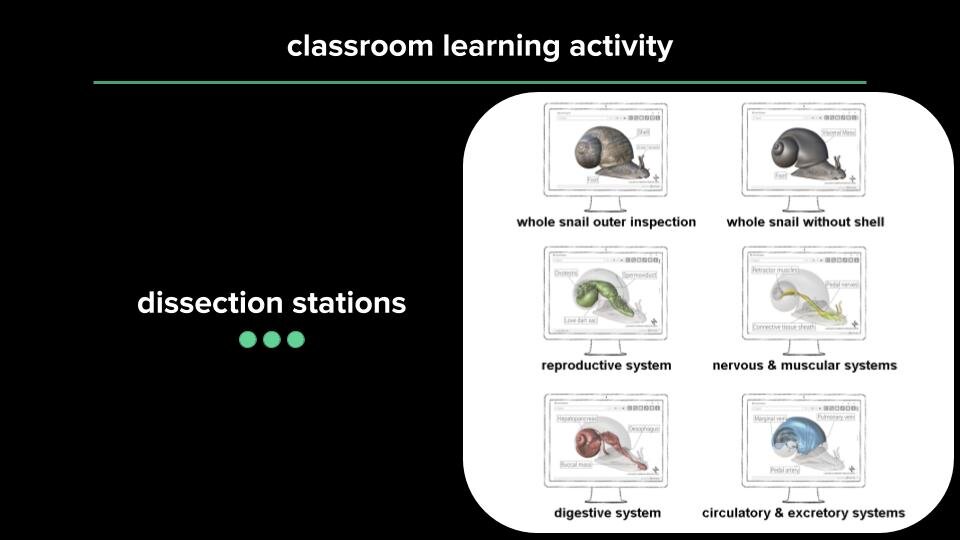
Slide 2: Our first teaching day was for an 8th grade biology class at Klenze-Gymansium Munich using both HAPI Helix and traditional dissection.
Slide 3: Learning stations were divided by body systems and had pre-dissected organs set up alongside Roman Snail Explorer Software.
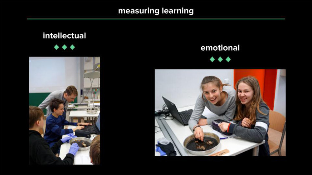
Slide 4: We measured both intellectual and emotional learning.
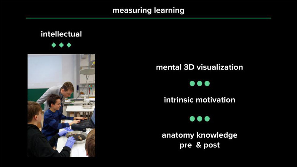
Slide 5: Intellectual learning components measured.
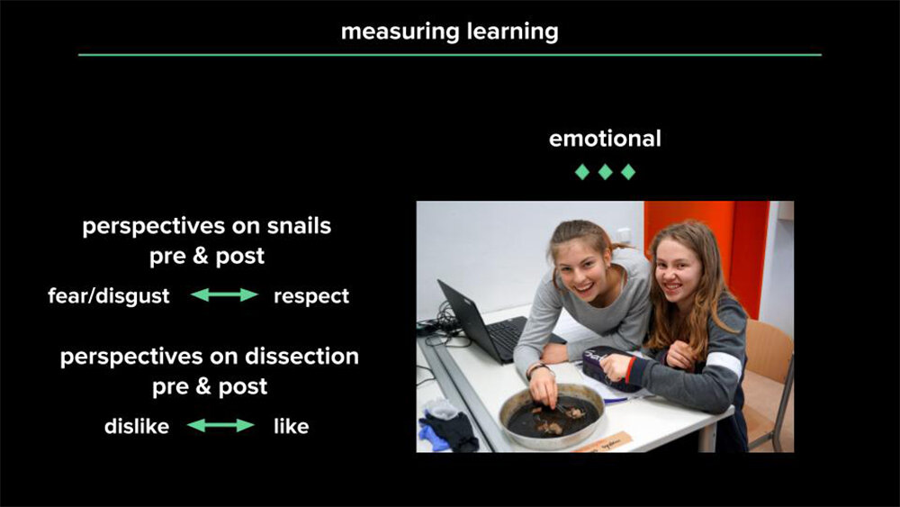
Slide 6: Emotional learning components measured.
All photos were taken by Bernhard Ruthensteiner, 7 March 2018 at Klenze Gymnasium Munich.

Roman Snail Explorer Software was also tested by >400 Biology Students at Ludwig-Maximilians University Munich as a supplement to snail dissection in the Introductory Zoology Course.
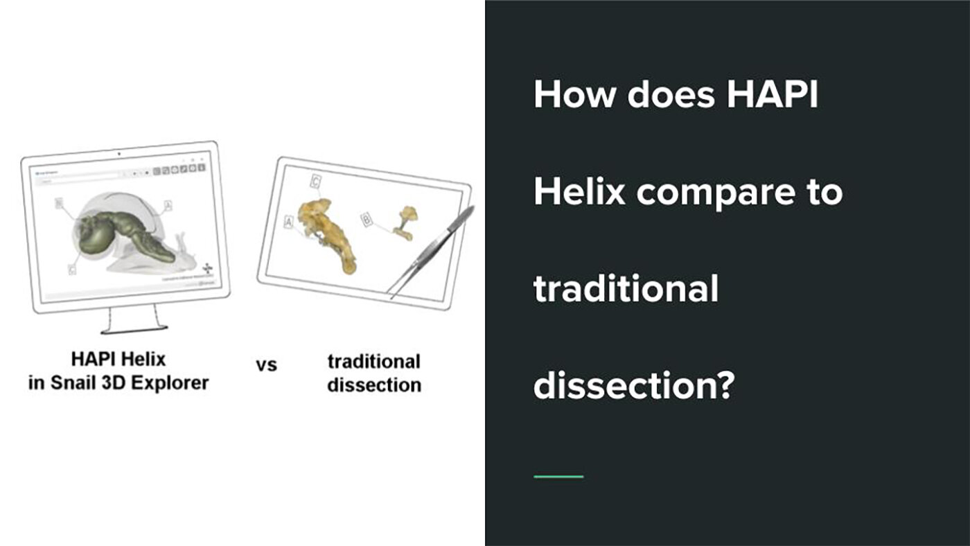
How does HAPI Helix compare to traditional dissection?
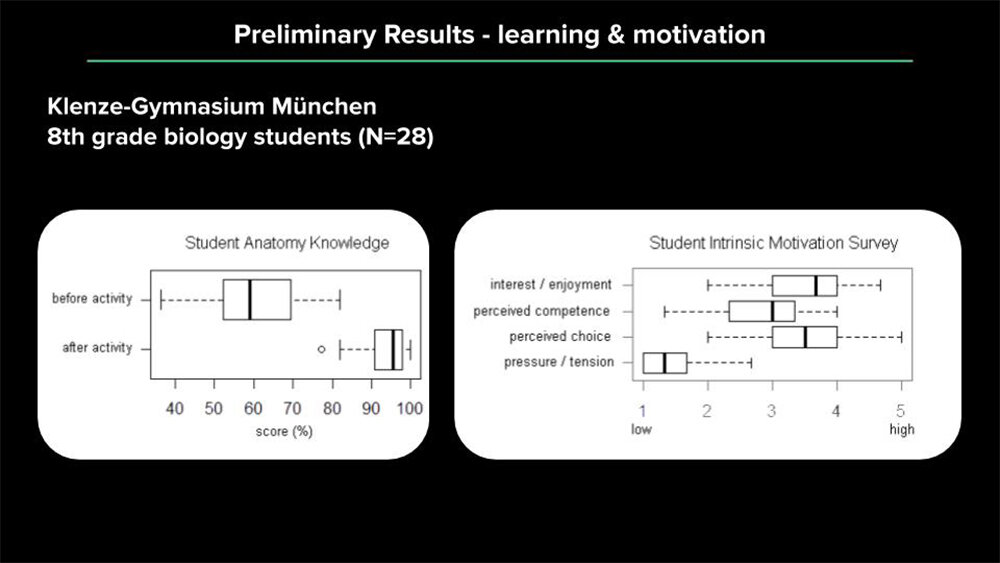
Slide 2: Students anatomy knowledge increased after the learning activity and overall enjoyment/motivation were high.
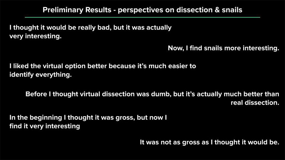
Slide 3: Quotes from university and 8th grade students on their experience using Roman Snail Explorer (translated from German).
More testing with students only using HAPI Helix or Traditional Dissection are needed to evaluate its effectiveness as a dissection alternative.


Check out HAPI Helix for yourself
Play around with interactive demos of the shell, body, and reproductive system.
Fun Facts About Snails
Stomach in your brain? A heart full of pee? Poop on your head? Snails are full of idiosyncrasies. Join me as I share the fun, weird facts I learned while working on my Master's thesis project.
Acknowledgements
A big thanks to everyone who came together and made this project possible, including:
My supervisors Bernhard Ruthensteiner, Lena von Kotzebue, Michael Apel, Gerhard Haszprunar and Michael John Gorman at the Bavarian State Collections of Zoology, Ludwig-Maximilians University (Departments of Biology and Didactics of Biology), Museum of Man and Nature (Museum Mensch und Natur Muenchen), and BIOTOPIA Natural History Museum (BIOTOPIA Naturkundemuseum Muenchen)
The folks at Effigos, AG (especially Jens Grosche, Thomas Barth, and Markus Schlaf)
Michael Troester, Martina, and the 8A students at Klenze-Gymansium München
Martina Bryce, Philomena Bodensteiner, Michael Bögle, Christina Schulte, Timea Neusser, Kathi Jörger, Matthias Starck, Carolin & Joachim Haug, and the Fall 2018 Introduction to Zoology students at Ludwig-Maximilians University Munich
My colleages at the Bavarian State Collections of Zoology (especially Peter Kohnert, Bastian Brenzinger, Michael Schroedl, Christina Laibl, Bruno Cancian, Jerome Moriniere, Rene Taenzler, Laura Hardulak, Alexander Cerwenka, Eva Lobbe-Bensch, and Mark Scherz)
Eike Neubert at the Natural History Museum of Bern
The German Academic Exchange Service (DAAD) for funding my Master’s studies
Lehre@LMU for funding the presentation of my thesis results at the Association for Science Teacher Education 2018 Conference in Baltimore, MD (USA) and carrying out the first application of my project
LMU Dual Career services for funding part of the collaboration between ZSM and Effigos AG to develop Snail 3D Explorer Software