3D pollination biology of cacao and its relatives
The first results of this project are now available in an article in Applications in Plant Sciences! Click here to give it a read and be sure to check out the whole issue, we made the cover image.

Looking for Cacao flowers in Fairchild Tropical Botanic Garden’s Tropical Plant Conservatory and Rare Plant House.
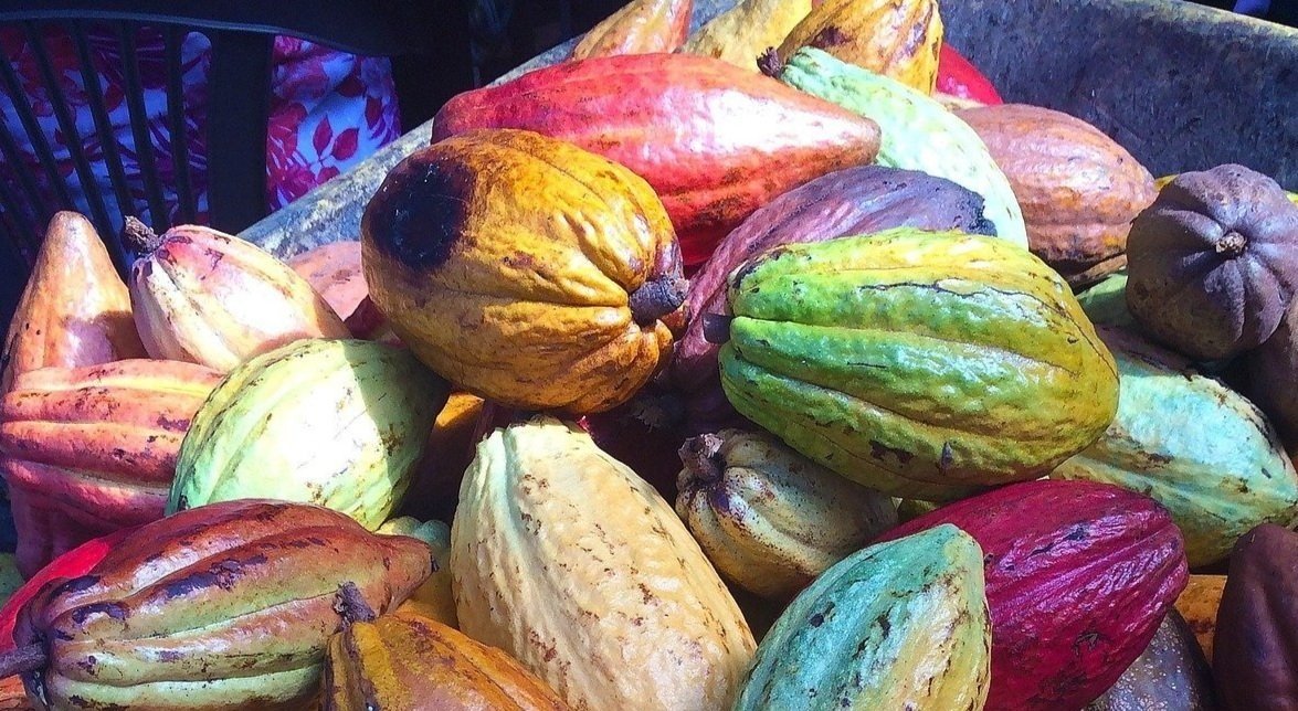
Cacao fruits image by myfriendkarla is licensed for public use by Pixabay.
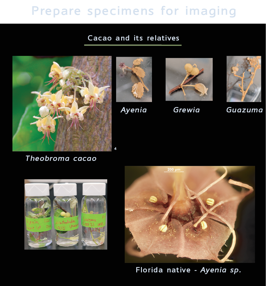
Despite the economic importance of chocolate ($182 billion global market predicted by 2025) and that insect pollinators have been shown to limit Cacao crop yields, little is known about the highly specialized pollination biology of Cacao, Theobroma cacao L., and its relatives, Ayenia sp. and Guazuma sp. (Report 5138783 2020, Toledo-Hernández et al. 2017).

Main suspects
The most likely suspects for Cacao’s pollinators are small flies (Diptera: Ceratopogonidae, Chloropidae, Phoridae), wasps (Hymenoptera: Eulophidae, Platygastridae), and thrips (Thysanoptera)
For pollination, insects must go through Cacao’s bizarre petals (petal sacs) to come into contact with the pollen-containing anthers. Then, they need to either escape the petals and fly to another flower (for cross pollination) or come into contact with the pollen-receiving stigma (for self pollination). Acquiring and depositing pollen via movement through these intricate 3-dimensional structures is size- and geometry-specific. For example, if an insect is too small or not the right shape, it could crawl through the flower without interacting with the anthers or stigma at all. Ceratopogonid midges (Diptera) are often named Cacao’s main pollinator, but field studies have found higher visitation rates for other small dipterans (Chumacero de Schawe et al. 2018). It is also possible that different insects are responsible for population across Cacao’s range. To determine the functional size limits of Cacao’s pollinators, 3D measurements of floral structures can be used and compared with suspected pollinator dimensions to make hypotheses on which floral visitors are most likely to be successful.

"Theobroma cacao" by Eric Hunt on Flickr is licensed under CC BY-NC-ND 2.0
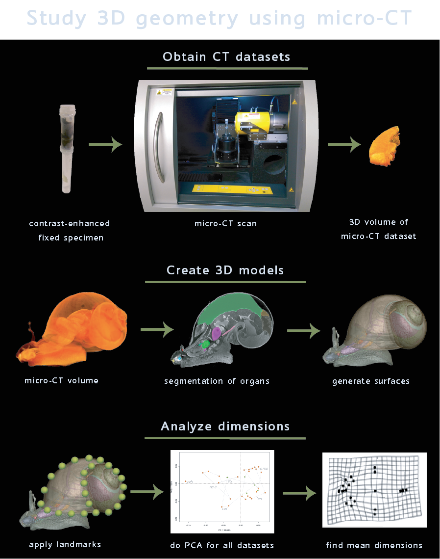
To precisely quantify plant-pollinator geometry and functional size limits of an effective pollinator for Cacao and its relatives, microcomputed tomography (micro-CT) and 3D geometric morphometrics (taking many measurements for individual flowers and comparing across species) can be used. After quantifying the functional size limits to evaluate Cacao’s suspected pollinators, the 3D datasets and measurements can be repurposed to develop a computational tool to expedite future 3D morphology studies on biological organisms.
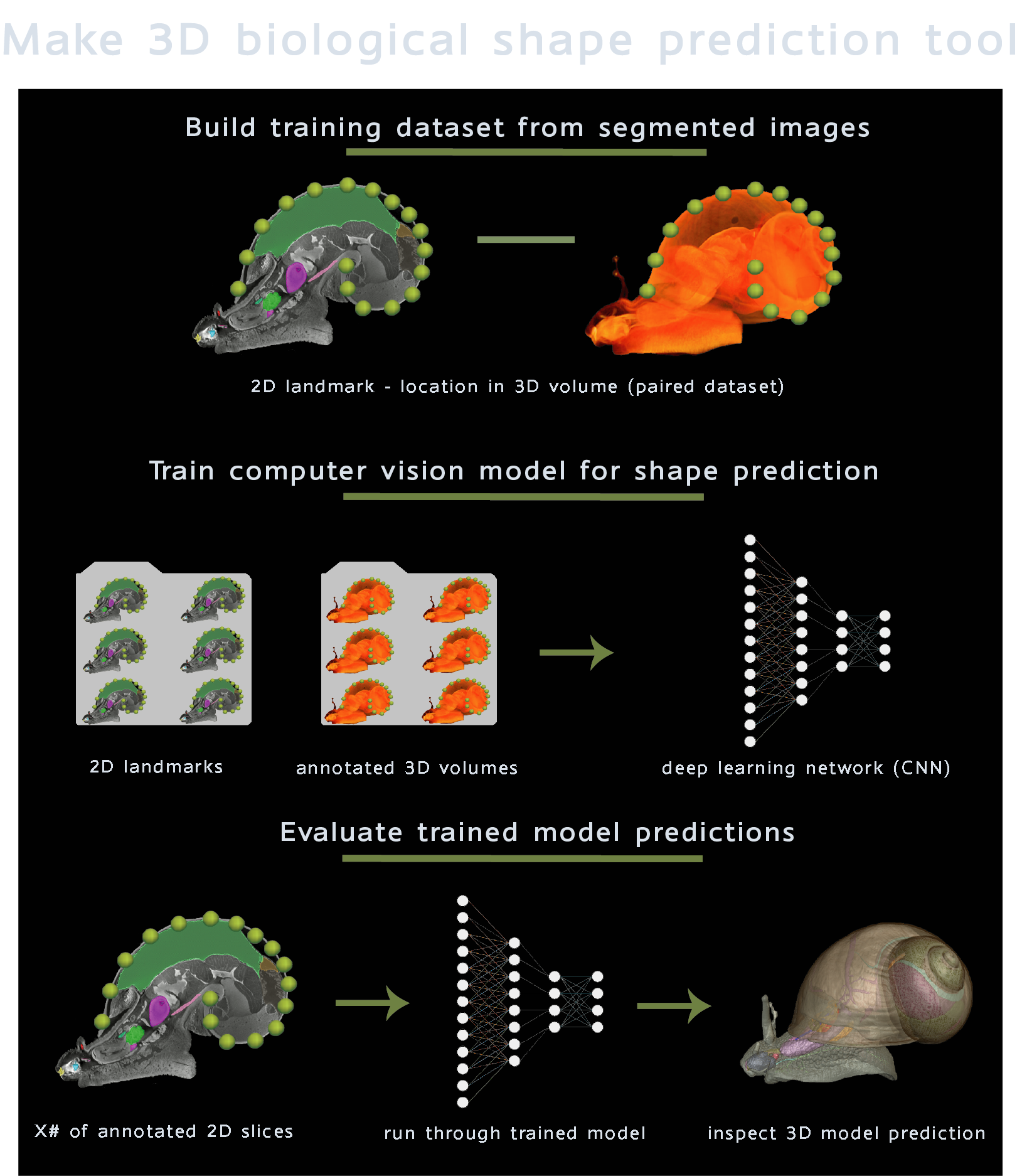
Computer vision has shown promise for expediting micro-CT dataset processing, visualization, and landmark placement, but applications to non-biomedical systems are just beginning (Porto and Voje 2020). For any machine learning pipeline, preparation and inspection of training data (which is used to teach the computer to identify patterns) is the most time-consuming step. By repurposing the datasets generated in the comparative morphology portion of our study, we will be able to save time on this crucial step to focus our efforts on refining 3D shape prediction algorithms that can save time and money for natural history museum digitization efforts by semi-automatically generating 3D models of specimens from raw 3D datasets.
Project Plan
For my PhD project, I will
(i) investigate functional size limits of Cacao flowers and their pollinators using micro-CT datasets of flowers and pollinators
(ii) repurpose obtained datasets to train computer vision models for landmark prediction
Understanding the diversity of life on earth has long relied on time-consuming methods to document and compare organisms through a combination of microscopy and dissection by taxonomic specialists. With advances in 3D imaging (especially micro-CT) and machine learning, large-scale comparative biology efforts are able to quantify and interpret patterns of biodiversity beyond what one taxonomist can accomplish.
Not only will this be the first study to obtain micro-CT images of Cacao, its relatives, and their pollinators, but it will also be the first use of computer vision for 3D landmark estimation in a non-biomedical context. Establishing functional size boundaries in Cacao pollination will facilitate identifying Cacao’s true pollinators and our findings can be built on by future ecological and behavioral field studies, advancing tropical biology and agroscience.
References
Chumacero de Schawe et al. 2018. Abundance and diversity of flower visitors on wild and cultivated cacao (Theobroma cacao L.) in Bolivia. Agrofor. Syst.
Porto and Voje 2020. ML-morph: A fast, accurate and general approach for automated detection and landmarking of biological structures in images. Methods Ecol. Evol.
Report 5138783 2020. Global Chocolate Market - Forecasts from 2020 to 2025. Research and Markets.
Toledo-Hernández et al. 2017. Neglected pollinators: Can enhanced pollination services improve cocoa yields? A review. Agric. Ecosyst. Environ.
Acknowledgements
Thanks to all our collaborators that are making this project possible!
My advisors Barbara Whitlock and Stefan Wuchty at University of Miami Departments of Biology and Computer Science
Funding for my graduate studies with my UM Fellowship from The University of Miami Graduate School and from the Christiane Tyson Research Fund for the Whitlock Lab’s 3D workstation
Felix Melian, Omar Gilzean, Luis Vidal, and Diego Rodriguez for assistance with purchasing and setting up the Whitlock Lab’s 3D workstation.
Sadegh Tale and Ali Ghahremaninezhad at University of Miami Department of Chemical, Environmental and Materials Engineering (CEME) for assistance and use of the Bruker Skyscan 1273
Brett Jestrow at Fairchild Tropical Botanical Gardens for help with specimen collection
Edward Leo Stanley and Gary Scheiffele at University of Florida and Florida Natural History Museum for help with use of the ZEISS Versa 620 XRM